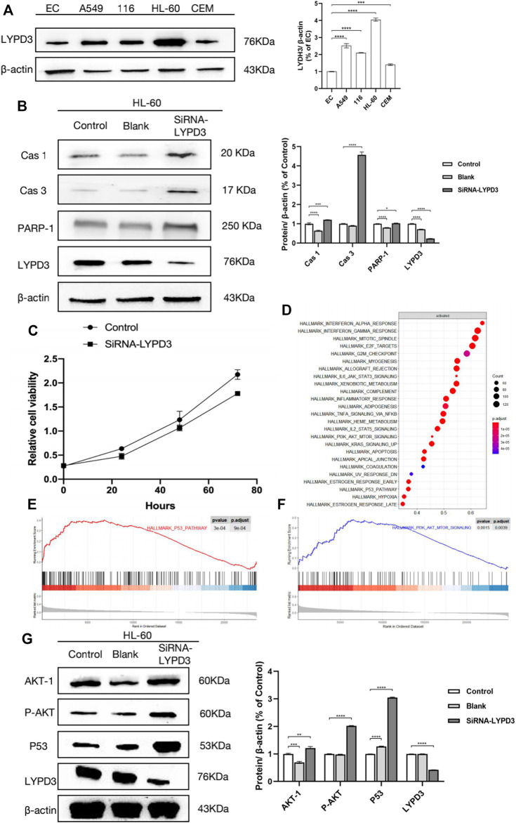
FIGURE 2.
(A) Expression of LYPD3 in cancer lines (the expression of LYPD3 is the highest in HL-60 cells); (B) LYPD3 gene knockdown mediated by siRNA-induced apoptosis in AML cells (HL-60 cells); (C) LYPD3 gene knockdown mediated by siRNA-suppressed proliferation in AML cells (HL-60 cells); (D) significantly enriched pathways in AML samples with high LYPD3 expression; (E) significantly enriched pathways (the P53 signaling pathways); (F) significantly enriched pathways (PI3K_AKT signaling pathway); (G) relationship between LYPD3 and the molecules Akt and P53 (the expression of the LYPD3 gene knockdown-mediated SiRNA group was obviously increased in p53 and PI3K_AKT signaling).

