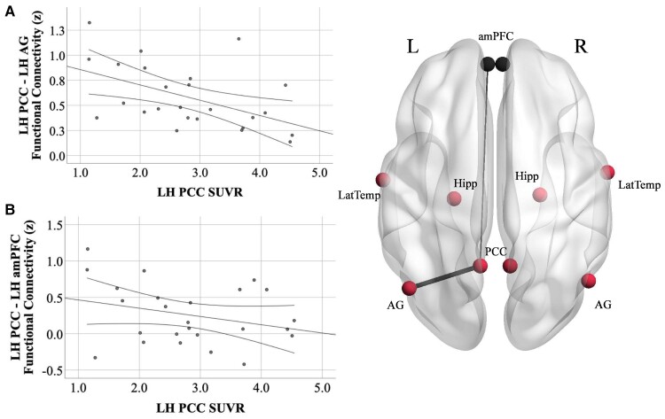
Figure 4.
Tau PET SUVR in the PCC is related to PCC-to-AG hypoconnectivity but not PCC-amPFC connectivity. (A) Increased tau PET signal in the left PCC is related to reduced functional connectivity between the left PCC and AG (r = −0.51, P = 0.01) in Aβ+ Alzheimer’s disease. (B) There was no relationship between tau PET signal in the left PCC and functional connectivity between the left PCC and amPFC (r = −0.29, P = 0.2). PCC, posterior cingulate cortex; AG, angular gyrus; amPFC, anterior medial prefrontal cortex; LH, left hemisphere. Functional connectivity units are Fisher’s z-scores. 95% confidence intervals are displayed. Results from the left hemisphere are shown here for illustrative purposes; complete results from both hemispheres are described in the text.

