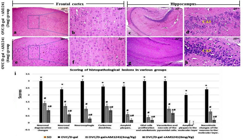
Fig 5.
H&E stained photomicrographs of frontal cortex and hippocampus areas of AD-model rats which treated with AM1241, (a-d) 3mg/kg and (e-h) 6mg/kg showing; few cerebral cortical scattered degenerated and necrotic neurons, absence of amyloid plaques with hippocampal disappearance of the amyloid plaques, mild degenerative changes of the DG and CA neurons. (i) The scoring of the observed histopathological lesions in various groups (Data are presented as median (n = 5 rats/group) using Kruskel-Wallis test followed the Mann-Whitney U test. Significantly different was considered at P<0.05, where; * significantly different when compared to control group, # significantly different when compared to OVC/D-gal group, ± significantly different when compared to AM1241 (3mg/Kg). Figs 5B-D and G-H are excluded from this article’s CC BY license. See the accompanying retraction notice for more information.

