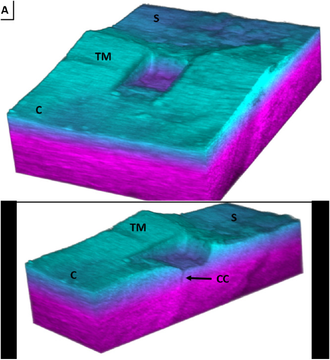
Figure 5.

A 3D spectral domain OCT reconstruction of an FLT channel (c-cornea, s-sclera, TM-trabecular meshwork, cc-collector channel). (A) Full image showing the wedge shape and well-defined edges of the FLT channel. (B) Cross-section of the FLT channel showing a collector channel.
