
Figure 2.
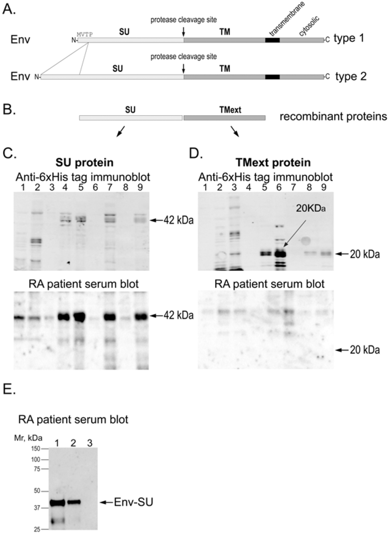
RA patient autoantibodies recognize the SU portion of HERV-K Env. (A) Schematic representation of type 1 and type 2 Env proteins. (B) The used recombinant SU and extracellular portion of TM (TMext) proteins from HERV-K_Xq21.33. (C) E. coli lysates expressing the SU portion of HERV-K Env protein immunoblotted with anti-6xHis tag antibody (upper panel) or RA patient serum (lower panel). (D) E. coli lysates expressing the extracellular region of the TM portion of HERV-K Env immunoblotted with anti-6xHis tag antibody (upper panel) or RA patient serum (lower panel). (E) Immunoblot of 1 μg (lane 1), 500 ng (lane 2), or 0 ng (lane 3) of purified HERV-K Env-SU with RA patient serum. The weaker lower band in lane 1 is a fragment of SU. E. coli: Escherichia coli; Env: envelope; HERV-K: human endogenous retrovirus K; RA: rheumatoid arthritis; SU: surface; TM: transmembrane.