. 2022 Mar 14;11:e76107. doi: 10.7554/eLife.76107

Figure 4. γ-Glutamyl phosphate reductase (GPR) domain ligand-bound mode and its conformation.

(A) The cryo-electron microscopy (cryo-EM) density of the GPR dimer structure and cartoon model is represented as an unliganded state in the P5CSGlu filament (green). (B) GPR dimer structure of the G5P-binding state in the P5CSGlu/ATPγS filament (coral). The conformation of the G5P-binding pocket and G5P (orange) is shown as stick representation. (C) GPR dimer structure of the NADP(H)-binding state in the P5CSMix filament (blue-violet). The conformation of the NADP(H)-binding pocket with NADPH (cyan) is shown as stick representation. (D) GPR dimer structure of the NADP(H)-released state in the P5CSMix filament (yellow). (E) Structural differences in the G5P-binding state (coral) and NADP(H)-binding state (blue-violet) of the GPR domain. Ligands are colored as in (B, C). (F) Superimposition of either the NADPH-binding domain (NBD) or the Rossmann-fold of the GPR domain at the G5P-binding state and NADP(H)-binding state using a single protomer.

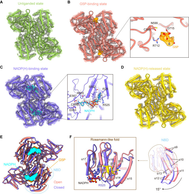
Figure 4.

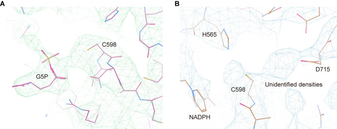
Figure 4—figure supplement 1. Representative cryo-electron microscopy (cryo-EM) densities for the active site of the γ-glutamyl phosphate reductase (GPR) domain.

Figure 4—figure supplement 1.

(A) Cryo-EM map quality of G5P ligand in the active site of the GPR domain in the P5CSGlu/ATPγS filament. (B) Unmodeled densities in the active site of the GPR domain at the NADP(H)-binding state, which may be the reaction product: Pi or GSA/P5C.
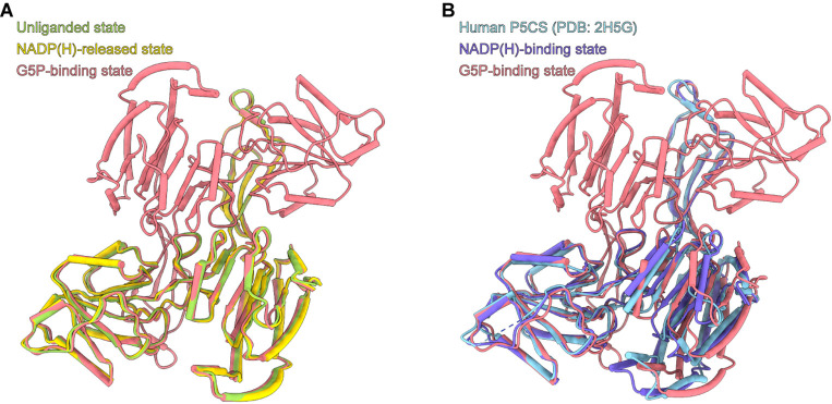
Figure 4—figure supplement 2. Comparison of the structures of the γ-glutamyl phosphate reductase (GPR) domain.

Figure 4—figure supplement 2.

(A) Superimposition of the GPR domain at the unliganded state (green), GPR domain at the NADP(H)-released state (yellow), and GPR domain dimer structure at the G5P-binding state (coral). (B) Superimposition of the GPR domain at the NADP(H)-binding state (blue-violet) and GPR domain dimer structure at the G5P-binding state (coral). Compared with the GPR domain of Drosophila P5CS at the NADP(H)-binding state (blue-violet), the GPR domain of human P5CS (cyan, PDB: 2H5G) has 56.74% sequence identity and root-mean-square-deviation (RMSD) value of 1.531 Å (398 atom pairs).
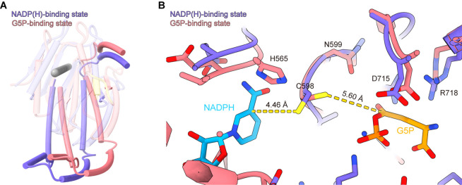
Figure 4—figure supplement 3. The NADPH-binding domain (NBD) rotation and view of the active site of the γ-glutamyl phosphate reductase (GPR) domain with its substrate.

Figure 4—figure supplement 3.

(A) The position of cylinder axes (gray) around which the NBD rotates. (B) Superimposition of the GPR domain at the G5P-binding state (coral) to that at the NADP(H)-binding state (blue-violet) on their active site; the distance between the ligands and catalytic residue C598 is shown by the dash line (yellow).
Figure 4—video 1. Structural transition of open and closed conformations of the γ-glutamyl phosphate reductase (GPR) domain.
Download video file (1.3MB, mp4)
Morph from the G5P-binding state (open conformation) to the NADP(H)-binding state (closed conformation) of the GPR domain.