. 2022 Feb 11;11:e72872. doi: 10.7554/eLife.72872

Figure 1. Dynein is required for acentrosomal pole focusing.

(A) Schematic representation of the Dynein auxin-inducible degron (AID) system for DHC-1 depletion in the C. elegans germ line. Methodologies for all three auxin treatments used in this study are depicted. (B) Immunofluorescence (IF) imaging of one-cell mitotically dividing embryos shows that auxin treatment causes efficient dynein depletion and canonical mitotic spindle defects (n = 22). Shown are tubulin (green), DNA (blue), ASPM-1 (red), and dynein (not shown in merge). (C) IF imaging of oocyte spindles in the Dynein AID strain shows that dynein is localized to the spindle, with increasing enrichment at acentrosomal poles at the anaphase transition; shown are tubulin (green), DNA (blue), dynein (red), and ASPM-1 (not shown in merge). Cortex is represented by the dashed line. (D) Representative images of oocyte spindles (GFP::tubulin and GFP::histone) in germline counting and corresponding quantifications; auxin treatment leads to splayed poles and spindle rotation defects. Cortex is represented by the dashed line. (E) IF imaging of Dynein AID conditions showing effects of 4 hr auxin treatment on metaphase (second row) and anaphase (bottom row); shown are tubulin (green), DNA (blue), ASPM-1 (red), and dynein (not shown in merge). ASPM-1 labeling supports initial observations of splayed poles seen in germline counting. (F) Quantifications of IF imaging shown in (E); meiotic spindles have significantly splayed poles upon auxin treatment. All scale bars = 2.5 µm. Controls for exposure to auxin plates can be found in Figure 1—figure supplement 1. Further experimentation with dynein mislocalization through aspm-1(RNAi) or lin-5(RNAi) can be found in Figure 1—figure supplement 2.

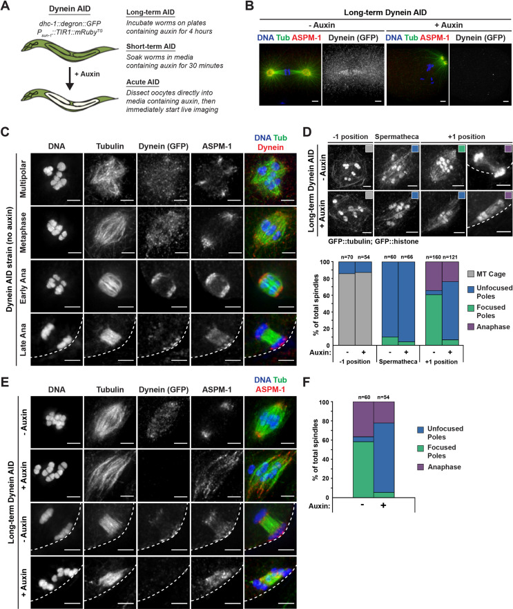
Figure 1.

Figure 1—figure supplement 1. Long-term treatment of wild-type worms with auxin does not have adverse effects on oocyte spindle assembly.

Figure 1—figure supplement 1.

Representative images of spindles (left side) assessed using germline imaging from a worm strain expressing GFP::tubulin, GFP::histone, and TIR1, but lacking degron-tagged DHC-1 (referred to as TIR1 control). Adult worms were either untreated or left on auxin plates for 4 hr, identical to our long-term Dynein auxin-inducible degron (AID) protocol. Three positions in the germline were analyzed to determine if auxin alone had adverse effects on spindle morphology; we observed no change in spindle phenotypes between control and auxin-treated worms. Cortex is represented by the dashed line. All scale bars = 2.5 μm.
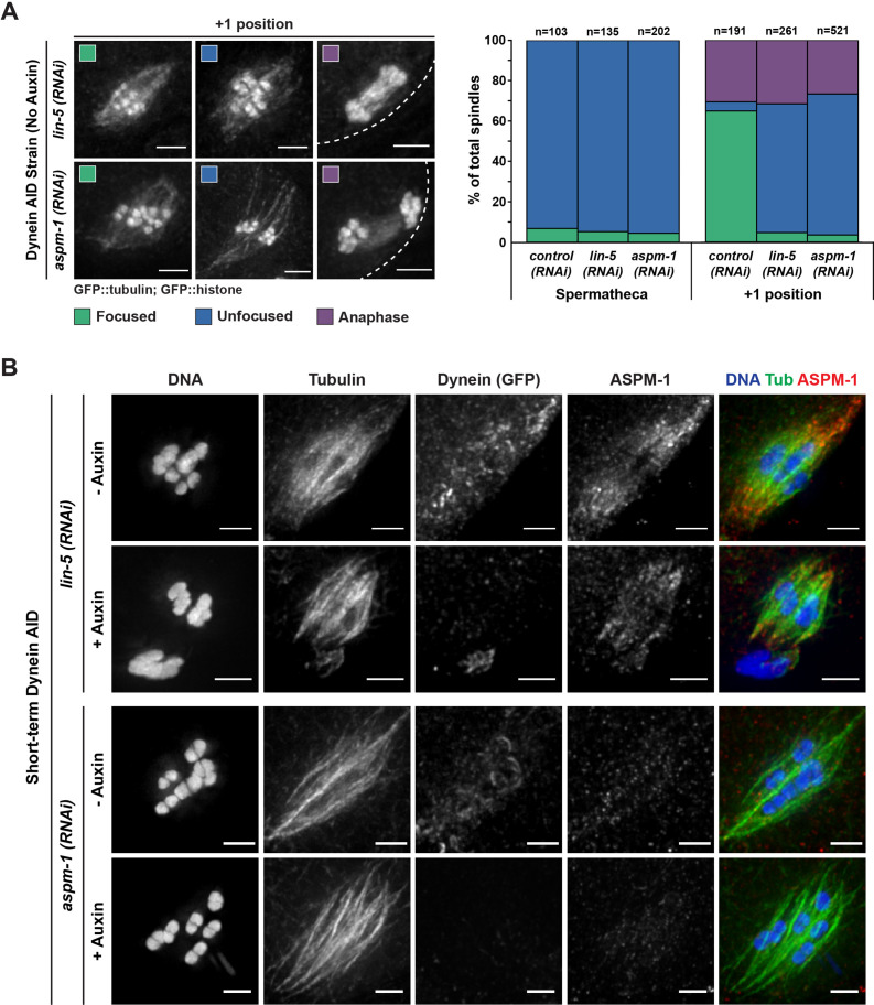
Figure 1—figure supplement 2. Oocyte spindles assembled following either ASPM-1 or LIN-5 depletion are morphologically similar to Dynein auxin-inducible degron (AID).

Figure 1—figure supplement 2.

(A) Representative images of spindles counted in germline imaging from the Dynein AID strain expressing GFP::tubulin and GFP::histone. In both aspm-1(RNAi) and lin-5(RNAi) conditions, splaying of acentrosomal poles can be observed in the +1 position. We also observed improper spindle rotation during anaphase, consistent with prior studies. Quantification of both aspm-1(RNAi) and lin-5(RNAi) revealed a drastic increase in unfocused poles at the +1 position and demonstrated similar ratios of unfocused poles as those seen in Dynein AID germline counting. Cortex is represented by the dashed line. (B) Immunofluorescence (IF) imaging of embryos from Dynein AID worms following either aspm-1(RNAi) or lin-5(RNAi); shown are tubulin (green), DNA (blue), ASPM-1 (red), and dynein (not shown in merge). ASPM-1 labeling further supports splaying of acentrosomal poles. Concurrent dynein depletion (rows 2, 4) does not appear to exacerbate the spindle defects observed in either aspm-1(RNAi) or lin-5(RNAi) alone. All scale bars = 2.5 μm.