
Figure 1.
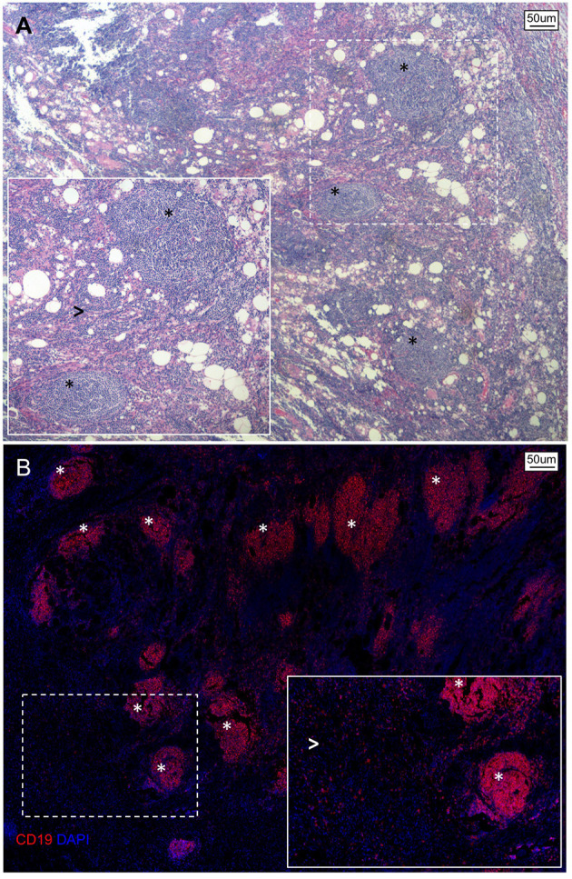
Spatial organization of B lymphocytes in pancreatic adenocarcinoma. (A) Hematoxylin/Eosin stain and (B) immunofluorescence staining of a representative tissue section of PDAC showing an abundant infiltrate of B lymphocytes organized as either multiple lymphoid structures (asterisks) or spread within the neoplastic lesion and tumor stroma (arrowheads).