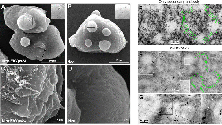
Figure 3.
Scanning and Transmission Electron Microscopy of transfected trophozoites. Trophozoites were treated as described for SEM or TEM. (A, B) SEM of trophozoites transfected (10,000x) with the pNeoEhvps23 (A) and pNeo (B) plasmids. Squared areas show part of the surface protuberance that are magnified at the right. Arrows: phagocytic cups. (C, D) Show part of the surface of trophozoites (25,000x). (E–G) TEM of thin sections of trophozoites transfected with the pNeoEhvps23. The helicoidal structures are remarked in green. (F, G) TEM images were obtained after treating the thin sections with α-EhVps23 followed by gold-labelled α-rat antibodies. Squared areas are magnified to show the very small vesicles in those regions. Arrowheads: label of the antibodies used to detect EhVps23. (F) The communication between vesicles where we visualized the presence of EhVps23 is shown. The magnification shows the communication channel between the vesicles.

