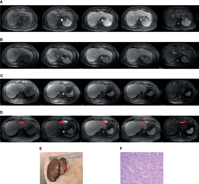
Figure 1.
Case examples of the non-HCC and positive HCC subgroups. Case 1 (A, B). MR images from a 49-year-old cirrhotic patient in the non-hepatocarcinogenic group. Axial fat-suppressed T1- and T2-weighted images show a cirrhotic liver morphology appears cirrhotic. Innumerable regenerative nodules are present throughout the liver, without suspicious nodules. (A) No HCC was found on the baseline MRI examination. (B) Similarly, no HCC lesion was seen after final follow-up at 70 months. Case 2 (C–F). MR images from a 34-year-old man in hepatocarcinogenic group. Axial fat-suppressed T1- and T2-weighted images show a cirrhotic liver with nodular contour. (C) Innumerable regenerative nodules were seen throughout the liver on baseline MRI examination. (D) At 16-month follow-up MRI, a new nodule about 3.1cm×2.0cm×2.0cm in size was clearly seen (red arrows) in segment four. The nodule appears hyperintense on T2-weighted image and hypointense on the pre-contrast T1-weighted images, with diagnostic enhancement of the arterial phase and subsequent washout on the portal venous and equilibrium phases. In addition, the tumor is surrounded by a fibrous capsule on the portal venous and equilibrium phases. (E) The liver explant specimen shows the hepatocellular carcinoma (red arrows) adjacent to Glisson’s capsule. (F) At histopathology (original magnification×40, hematoxylin-eosin staining), a moderately differentiated hepatocellular carcinoma is confirmed.

