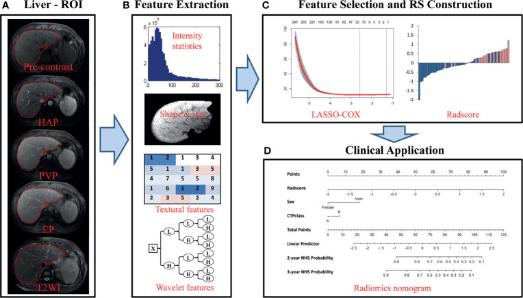
Figure 2.
Flowchart of the study. (A) Liver segmentation is performed on axial T2-weighted and contrast-enhanced T1-weighted imaging. Pre-contrast, pre-contrast phase; HAP, hepatic arterial phase; PVP, portal venous phase; EP, equilibrium phase; T2WI, T2 weighted imaging. (B) Four types of features are extracted from within the defined liver areas on the MR images, including quantify intensity, shape, texture and wavelet texture of the entire liver. (C) The LASSO method was applied to select suitable features and the selected features were used to calculate the Rad-score for the radiomic nomogram. (D) The radiomic signature incorporated the prognostic clinical risk factors in a final radiomic nomogram for individual evaluation.

