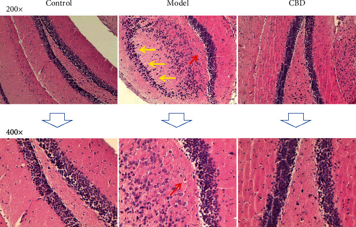
Figure 2.

Representative histopathological microphotographs of the substantia nigra area in the midbrain (HE-staining). Yellow arrows indicate obviously fading substantia nigra areas compared twith the control group, and red arrows indicate typical Lewy bodies.
