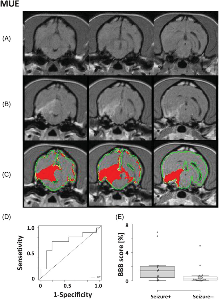
FIGURE 3.

Detection of blood‐brain barrier dysfunction (BBBD) in dogs with meningoencephalitis of unknown origin (MUO) using subtraction enhancement analysis (SEA). A‐C, Representation of MUO in a dog. A, T1‐weighted precontrast images; B, T1‐weighted postcontrast images; C, positive voxels as detected by SEA using the high threshold (HT, red) were superimposed on postcontrast T1‐weighted images. D, Receiver operating characteristic curve (ROC) analysis of BBBD scores was performed to determine the optimal cutoff point between MUO dogs with and without seizures. E, Box plot comparing BBB scores of MUO dogs with or without epileptic seizures. Center lines show the medians; box limits indicate the 25th and 75th percentiles as determined; whiskers extend to 1.5 times the interquartile range from the 25th and 75th percentiles (seizure+; n = 11, seizure−; n = 21)
