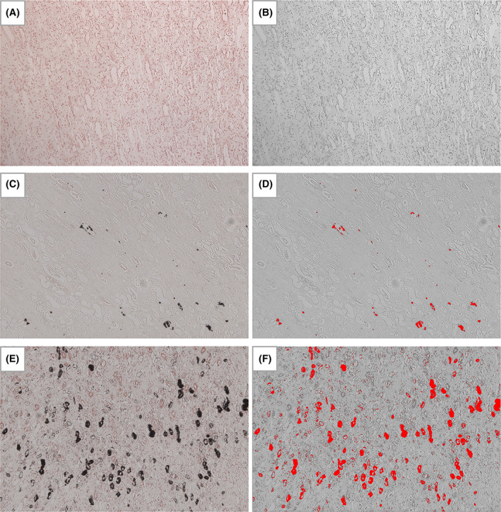
FIGURE 1.

Kidney, von Kossa stain, ×10 magnification. An example of each nephrocalcinosis grade (0‐2) is shown as follows: (A) and (B) cat no. 7 with grade 0 nephrocalcinosis (0.004%); (C) and (D) cat no. 34 with grade 1 nephrocalcinosis (0.327%); (E) and (F) cat no. 39 with grade 2 nephrocalcinosis (5.51%). Von Kossa positive staining is outlined in black coloring in the original images (A, C, and E), or bright red in color after processing using ImageJ (B, D, and F; 8‐bit color; threshold 0‐92) for the quantification of the proportional area of nephrocalcinosis in each case (n = 51)
