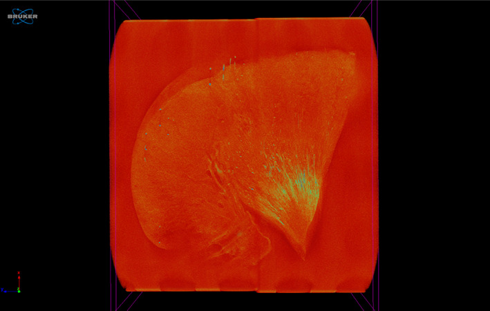
FIGURE 6.

Micro‐computed tomography (micro‐CT) image of a kidney section with grade 2 nephrocalcinosis (cat no. 17), projection images were reconstructed into tomograms and volume‐rendered 3‐dimensional visualizations were created using Bruker micro‐CT SkyScan software (NRecon and CTVox, respectively). Renal calcification is outlined in yellow‐green coloring
