. 2022 Mar 18;63(4):349–356. doi: 10.3349/ymj.2022.63.4.349

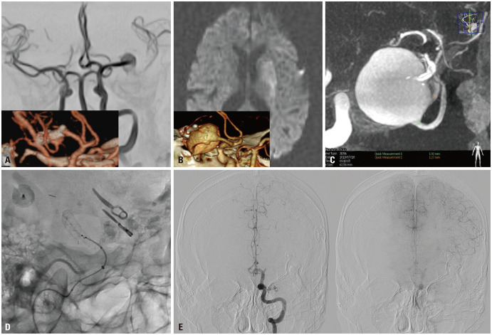
Fig. 1. Illustrative case. A 57-year-old male presenting with acute infarction on left basal ganglia. (A) In November 2003, the patient underwent surgical clipping for unruptured large aneurysm located at the left MCA. After the surgery, small neck remnant was observed on postoperative CTA (lower box). (B) In May 2019, the patient developed right hemiparesis with acute infarction on left basal ganglia on diffusion-weighted image, and follow-up CTA (lower box) showed major recurrence of the previously clipped aneurysm. (C) DSA showed a giant aneurysm involving MCA trifurcation and the inferior trunk had the largest diameter of about 1.92 mm. (D) We successfully deployed FRED from the inferior trunk to M1 without procedural complication in December 2019. (E) One month later, the patient had right hemiparesis again due to acute infarction on the left periventricular white mater. Follow-up DSA demonstrated left M1 occlusion with leptomeningeal collateral from the ACA. The aneurysm was completely occluded on DSA. ACA, anterior cerebral artery; CTA, computed tomography angiography; DSA, digital subtraction angiography; FRED, Flow Re-direction Endoluminal Device; MCA, middle cerebral artery.

Fig. 1