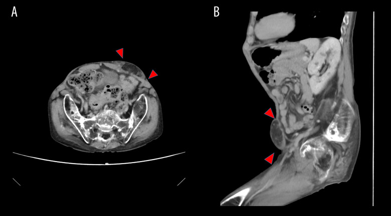
Figure 1.
Abdominal contrast-enhanced computed tomography scan showing a spigelian hernia in a patient with juvenile rheumatoid arthritis. (A, B) Abdominal contrast-enhanced computed tomography (CT) scan. Abdominal contrast medium-enhanced CT revealed a mass of low signal attenuation, which suggested the presence of mesenteric fat between the thin ventral muscles in the left-lower abdominal wall (red arrow).

