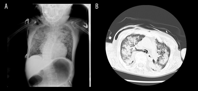
Figure 3.
Chest computed tomography scan of a patient with juvenile rheumatoid arthritis after difficult endotracheal intubation. (A) Chest radiograph after intubation showed pulmonary edema in both lobes and no cardiomegaly. (B) Chest computed tomography showed bilateral infiltration and diffuse ground-glass opacities, which were suggestive of pulmonary edema.

