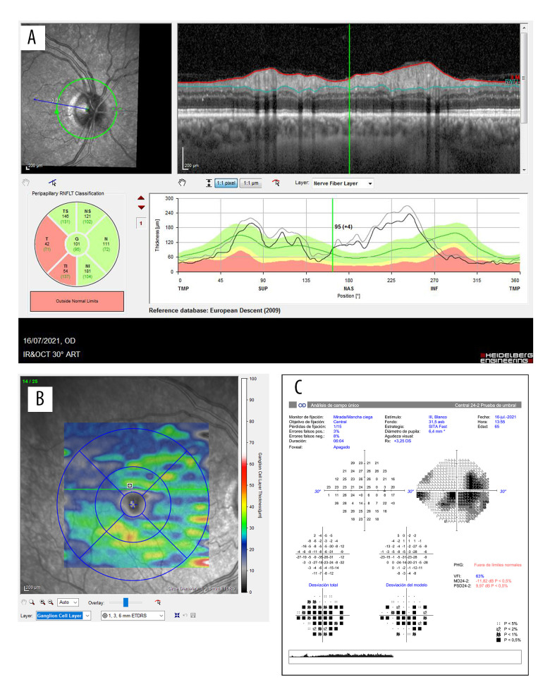
Figure 7.
Evolution of the OCT and Humphrey 24-2 visual field in the second case. A shows the RNFL of the right eye and the correspondence of the atrophic quadrants in the affected eye after a month of the symptoms. B illustrates general loss of ganglion cells of the right eye. Image C illustrates the Humphrey 24-2 visual field of the right eye, showing a centrocecal scotoma.

