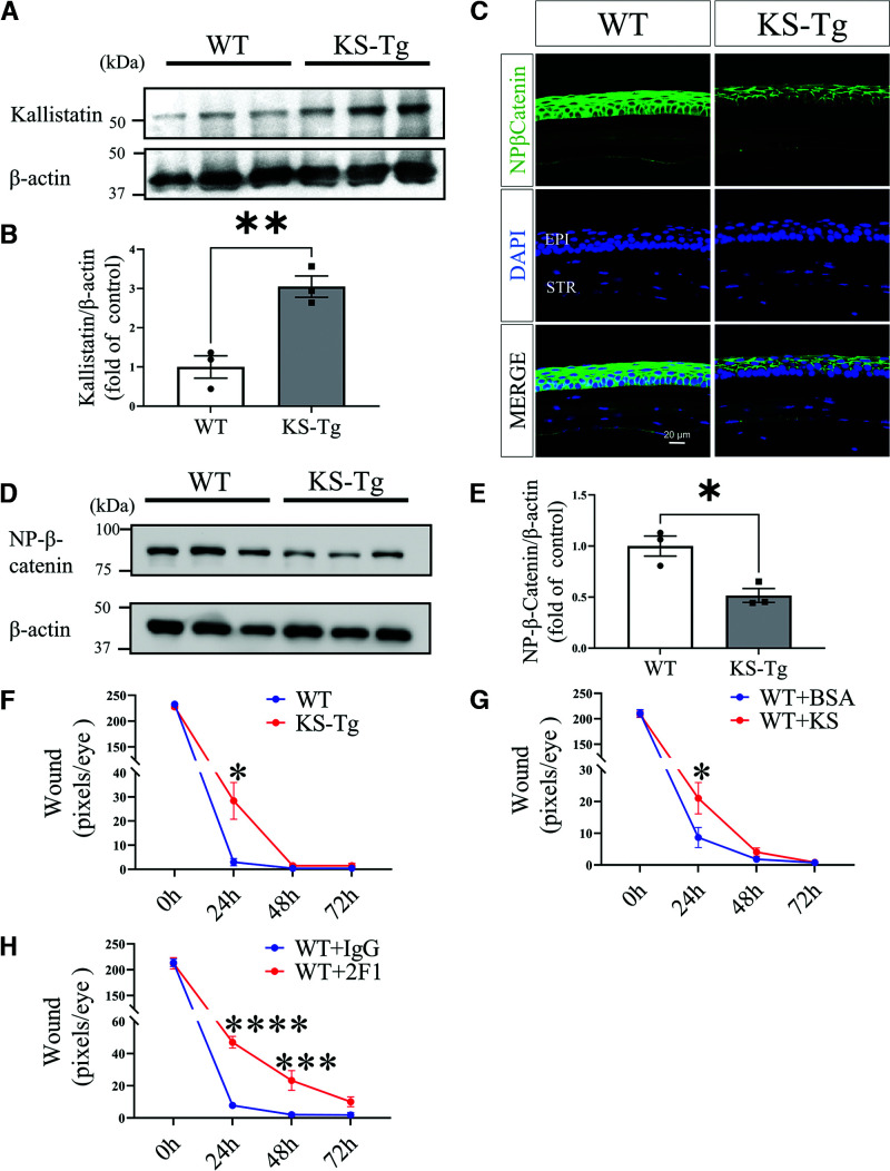
Figure 4.
Delayed corneal wound healing in KS-Tg mice by inhibition of the Wnt signaling pathway. A: Western blot analysis of kallistatin in the cornea of 5-month-old KS-Tg mice and WT littermates. B: Densitometry analysis of kallistatin in panel A and normalized by β-actin levels (n = 3). C: Representative images of nonphosphorylated β-catenin (NP-β-catenin) in the corneas of 5-month-old KS-Tg mice and WT littermates (n = 5). D: Western blot analysis of NP-β-catenin in the corneas. E: Densitometry analysis of NP-β-catenin in the cornea in panel D and normalized by β-actin levels (n = 3). F: The wound in the cornea from KS-Tg and WT mice was quantified after fluorescein staining using the pixel per eye with ImageJ (n = 10). G: Subconjunctival injection of kallistatin protein (KS, 10 µg/eye) into WT mice, with BSA for control. Wound severity was quantified at postwounding time as indicated (n = 10). H: WT mice received a subconjunctival injection of 10 µg of Mab2F1 with IgG for control, and then wound severity was quantified at the indicated time points (n = 8). All values are mean ± SEM. *P < 0.05; ***P < 0.001; ****P < 0.0001.

