Figure 8.
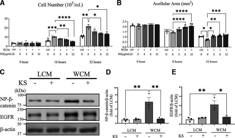
Effect of kallistatin on HCEC. A: HCEC were treated with 20% LCM or WCM, with or without different concentrations of human kallistatin, as indicated. Viable cells were quantified at 0, 16, and 32 h (n = 3–6). B: HCEC were treated with WCM and various concentrations of kallistatin after 100% confluence. An in vitro “wound” was created, and then 6 images of each scratch were captured at the indicated time points. The acellular area was measured by ImageJ (n = 5). C: Representative Western blots of nonphosphorylated β-catenin (NP-β-catenin) and EGFR in HCEC with indicated treatment. HCEC were treated with 20% LCM or WCM, with or without 12 µmol/mL kallistatin for 48 h. Protein levels of NP-β-catenin (D) and EGFR (E) in panel C were quantified by densitometry (n = 3). Values are mean ± SEM. *P < 0.05; **P < 0.01; ***P < 0.001; ****P < 0.0001.

