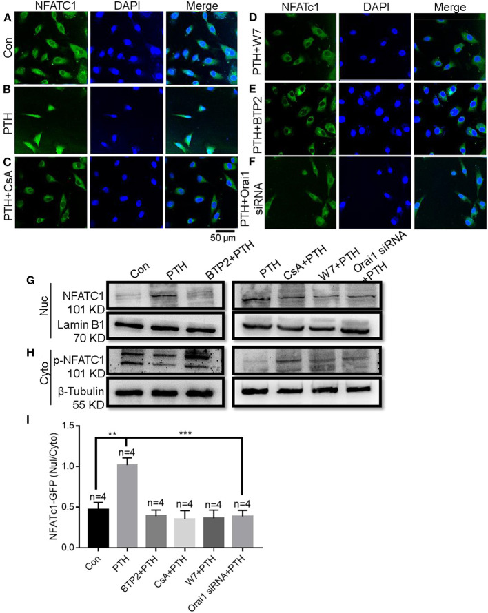
Figure 6.
Role of Orai1-mediated store-operated Ca2+ entry (SOCE) in parathyroid hormone (PTH)-induced NFAT nuclear translocation in human umbilical vein endothelial cells (HUVECs). (A–F) Representative confocal microscopy images and summary data (I) showing NFATC1 distribution in HUVECs treated for 24 h with (A) vehicle control, (B) 100 pM PTH, (C) PTH + CsA (calcineurin inhibitor), (D) PTH + W7 (calmodulin antagonist), (E) PTH + BTP2 (an Orai nonspecific inhibitor) or (F) PTH + Orai1 siRNA transfection. Green fluorescence indicates NFATC1; Blue, 4′,6-diamidino-2-phenylindole (DAPI) indicates nuclei. Data are shown as the mean ± SEM; n = 4. **P < 0.01, ***P < 0.001 vs. Con or PTH analyzed by one-way analysis of variance followed by Dunnett's multiple comparisons test. (G,H) Representative Western blot images showing fractionation assay results indicating the presence of p-NFATC1 in the cytoplasmic [Cyto, (H)] and NFATC1 in the nuclear [Nuc, (G)] extracts under the same treatment conditions as for confocal microscopy analyses. Lamin B1 is a nuclear marker; β-Tubulin is a cytoplasmic marker. (I) Summary data showing the ratio of green fluorescence intensity of NFATc-GFP in the nuclear (Nuc)/cytoplasmic (Cyto).

