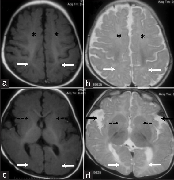
Figure 1.

MRI brain images of an 8-month-old child. Axial T1 (A) and T2 weighted images (B) at the level of the perirolandic region shows delayed myelination in the frontal (asterisk) and parietal (arrows) white matter bilaterally. In comparison to the normally myelinated perirolandic white matter, there is mild T2 hyperintensity and relative T1 iso to hypointensity in these regions. Axial T1 (C) and T2 weighted images (D) at the level of the basal ganglia shows similar changes at the level of the anterior limbs of internal capsule (dashed arrows) and calcarine sulcus white matter (white solid arrows). Bilateral open opercula (black solid arrows, D) also noted
