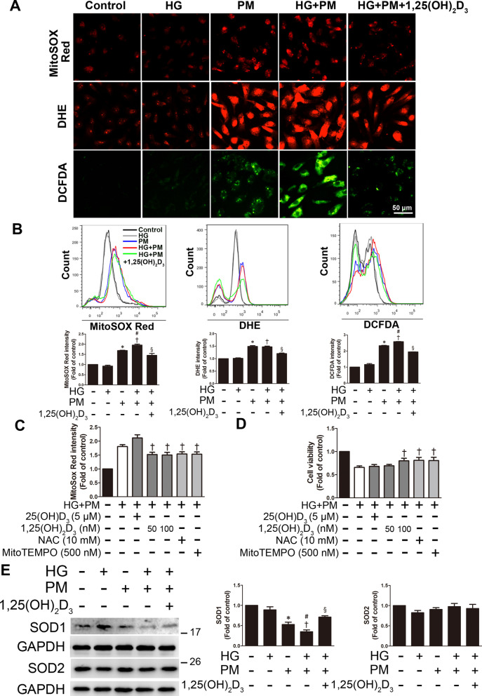
Fig. 2.
PM aggravated ROS production in endothelial cells treated with high glucose, and 1,25(OH)2D3 pretreatment attenuated this change. HUVECs were pretreated with high glucose (30 mM) for 24 h and then treated with PM (50 μg/mL) for 8 h. Before exposure to PM, HUVECs were pretreated with 100 nM 1,25(OH)2D3 for 12 h. A Mitochondrial and cellular ROS levels were detected by staining HUVECs with MitoSOX Red, DHE and DCFDA and examining them under a fluorescence microscope. Bar = 50 μm. B Mitochondrial and cellular ROS levels were detected by staining HUVECs with MitoSOX Red, DHE and DCFDA and analyzed using flow cytometry. C HUVECs were pretreated with 5 μM 25(OH)D3, 50 or 100 nM 1,25(OH)2D3, 500 nM MitoTEMPO and 10 mM NAC (an ROS scavenger) for 12 h and 1 h, respectively. Mitochondrial ROS was detected using MitoSOX Red and a flow cytometer. D HUVECs were pretreated with 5 μM 25(OH)D3, 50 or 100 nM 1,25(OH)2D3, 500 nM MitoTEMPO and 10 mM NAC for 12 h and 1 h, respectively. The MTT assay was used to evaluate cell viability. E The levels of SOD1 and SOD2 expression were detected using Western blotting. *P < 0.05 compared with the control group; †P < 0.05 compared with the HG group; #P < 0.05 compared with the PM group; §P < 0.05 compared with the HG + PM group

