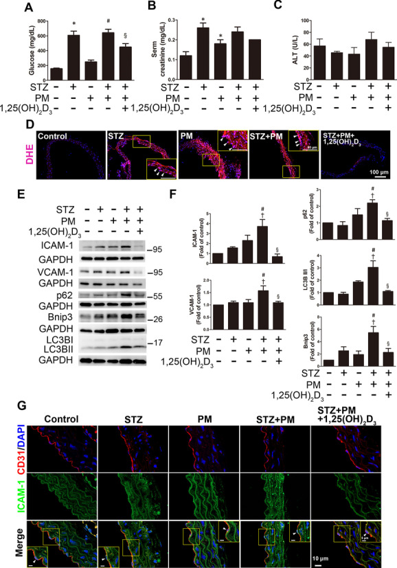
Fig. 7.

The effects of 1,25(OH)2D3 on mouse aortas under high glucose and PM conditions. Diabetes was induced in mice by intraperitoneally injecting STZ (55 mg/kg). Mice received PM (10 mg/kg) via intratracheal injection under anesthesia to simulate exposure to air pollution. 1,25(OH)2D3 was administered at a dose of 7 μg/kg per day for two weeks. The levels of A blood glucose, B serum creatinine and C ALT were analyzed. D DHE staining showed the level of oxidative stress in the mouse aorta. Red fluorescence in sections incubated with DHE indicated O2•− production; blue fluorescence in sections stained with DAPI indicated the cell nucleus. Endothelial cells are indicated with white arrowheads. Bar = 100 μm. E, F The expression levels of ICAM-1, VCAM-1, p62, LC3B, and Bnip3 were detected using Western blotting. G The colocalization analysis of immunohistochemical staining showed that the expression of ICAM-1 in endothelial cells (positive for CD31) in the STZ + PM group was stronger than that in control mice. Yellow boxes show the magnification of endothelial cells stained with ICAM-1. Red fluorescence in sections indicated CD31; green fluorescence in sections indicated ICAM-1; blue fluorescence in sections indicated the cell nucleus. White arrowheads indicated the endothelial cells. Bars = 5 or 10 μm as indicated in the panels. *P < 0.05 compared with the control group; †P < 0.05 compared with the STZ group; #P < 0.05 compared with the PM group; §P < 0.05 compared with the STZ + PM group
