Fig. 1.
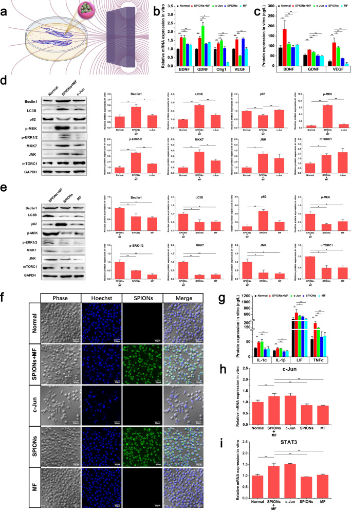
SPION-mediated magnetic actuation induces repair phenotypes in RSC96 cells in vitro. a shows the MF environment used in cell experiments in vitro. RSC96 cells coincubated with SPIONs and were magnetized by ingestion of SPIONs. One perpetual cuboid neodymium magnet provided approximately 6.0 T/m of MF to the RSC96 cells at the center of the dish. b The relative mRNA expression of regeneration-related neurotrophic factors in different experimental groups was detected through qRT-PCR. c The protein expression of regeneration-related neurotrophic factors in different experimental groups was detected by ELISA. d The protein expression levels of autophagy markers and regeneration-related signaling pathway biomarkers between the Normal, SPIONs + MF and c-Jun groups were detected through WB. e The protein expression levels of autophagy markers and regeneration-related signaling pathway biomarkers between the SPIONs + MF, SPIONs and MF groups were detected through WB. f The morphology of RSC96 cells in different experimental groups was observed by confocal laser scanning microscopy. g The protein expression of immune-related cytokines in different experimental groups was detected by ELISA. h, i The relative mRNA expression of Schwann cell repair phenotype-related transcription factors (c-Jun and STAT3) in different experimental groups was detected by qRT-PCR. Each experiment was carried out in triplicate. The relative mRNA expression was calculated by using the 2-ΔΔCT method. The values are represented as the mean ± SD. Scale bar = 30 µm in panel f. *P < 0.05, **P < 0.01

