Fig. 3.
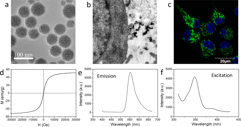
Synthesis and characterization of fluorescent-magnetic bifunctional SPIONs. a The high resolution transmission electron microscopy (HRTEM) image shows that SPIONs have an uniform size distribution, with a Fe3O4·Rhodamine 6G SPs core size of 50 nm and a thin PDA shell of about 6 nm. b TEM image shows the successive stage of SPIONs internalization, which suggest that SPIONs has excellent biocompatibility and neuronal affinity. c CLSM image of RSC96 cells after incubation with SPIONs exhibit a strong green fluorescence, which demonstrates that the SPIONs possess an ideal capacity to magnetize Schwann cells. d Magnetic hysteresis curve of SPIONs shows the saturation magnetization (Ms) is 51 Am2/kg and without any evident remanence or coercivity, suggesting the superparamagnetic property of SPIONs. Photoluminescence spectra of SPIONs show optimal emission (e) and excitation (f) wavelengths at around 556 and 350 nm

