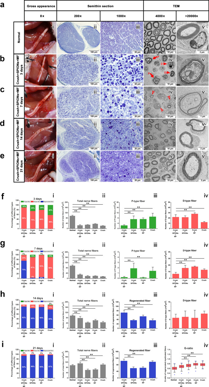
Fig. 6.
SPION-mediated magnetic actuation promotes morphological regeneration of the sciatic nerve. At 3, 7, 14, and 21 days after sciatic nerve crush injury, the gross appearance, semithin sections and ultrathin sections were observed by stereomicroscopy, optical microscopy, and TEM. The morphology and microstructure of nerves in different experimental groups were observed and quantitatively assessed. a The gross appearance (i) of the sciatic nerve immediately after crush injury and the microstructure of the normal nerve (ii–v). b In the magnetic actuation (Crush + SPIONs + MF) group, the gross appearance (i) and microstructure of the sciatic nerve at 3 days after crush injury were observed and showed obvious Wallerian degeneration (ii-v). c In the Crush + SPIONs + MF group, the gross appearance (i) and microstructure of the sciatic nerve (ii–v) at 7 days after crush injury were observed and showed new regenerated nerve fibers (iv, v). d In the Crush + SPIONs + MF group, the gross appearance (i) and microstructure of the sciatic nerve (ii-v) at 14 days after crush injury were observed, and the thickness of the myelin sheath of the regenerated nerve fibers was increased (iv, v). e In the Crush + SPIONs + MF group, the gross appearance (i) and microstructure of the sciatic nerve (ii-v) at 21 days after crush injury were observed, and the morphology of the regenerated nerve fibers returned to nearly normal (iv, v). f Three days after crush injury, the proportions of various nerve fibers (i), the number of total nerve fibers (ii), the number of P-type nerve fibers (iii), and the number of D-type nerve fibers (iv) of the sciatic nerve in the Crush + SPIONs + MF, Crush + SPIONs, MF, and Crush groups were quantitatively assessed. g Sciatic nerve morphometric assessment was performed in each of the four experimental groups 7 days after crush injury. h At 14 days after crush injury, the proportions of various nerve fibers (i), the number of total nerve fibers (ii), the number of regenerated nerve fibers (iii), and the number of D-type nerve fibers (iv) of the sciatic nerve in the four experimental groups were quantitatively assessed. i At 21 days after crush injury, the proportions of various nerve fibers (i), the number of total nerve fibers (ii), the number of regenerated nerve fibers (iii) and the G-ratio (iv) of the sciatic nerve in the four experimental groups were quantitatively assessed. The long red solid arrows indicate the P-type nerve fibers and the short red solid arrowheads indicate the D-type nerve fibers in panel b (iv), the short red hollow arrowheads indicate the regenerated nerve fibers in panel c (iv). *P < 0.05, **P < 0.01

