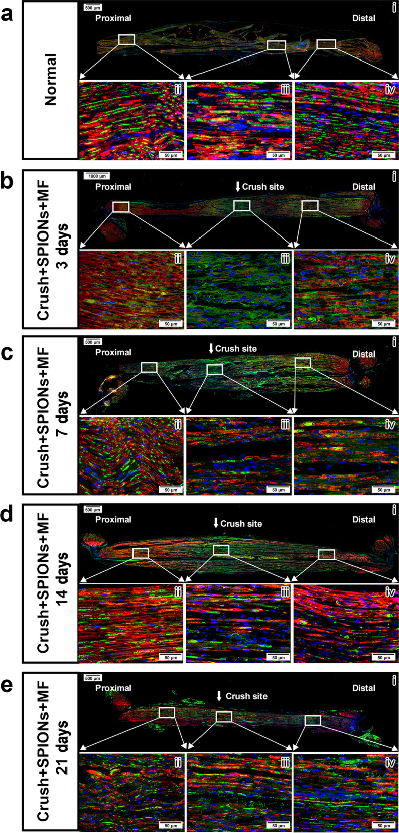
Fig. 7.

Regeneration and repair of the sciatic nerve after crush injury in the magnetic actuation group. The regeneration of axons (green) and the interaction of axons with Schwann cells (red) after crush injury of the sciatic nerve were observed by immunofluorescence staining using neurofilament heavy chain antibody and S100β antibody. a Microstructure of normal sciatic nerve. Schwann cells form myelin sheaths (red) around axons (green). b In the Crush + SPION + MF group, the microstructure of the sciatic nerve 3 days after crush injury showed obvious Wallerian degeneration at the crush site (iii) and distal end (iv). c In the Crush + SPION + MF group, the microstructure of the sciatic nerve 7 days after crush injury showed the growth of new regenerated axons at the crush site (iii), and the regenerated axons were not wrapped by the myelin sheath (iv). d In the Crush + SPION + MF group, the microstructure of the sciatic nerve 14 days after crush injury showed numerous regenerated axons at the crush site (iii), and significant remyelination was observed. Meanwhile, bands of Bungner (red) formed from Schwann cells arranged in rows were seen at the distal stump of the nerve (iv). e In the Crush + SPION + MF group, the microstructure of the sciatic nerve 21 days after crush injury showed robust axon regeneration at both the crush site (iii) and the distal end of the nerve (iv), and the regenerated axons obtained good remyelination. Proximal = proximal stump of sciatic nerve to the crush site. Distal = stump of sciatic nerve distal to the crush site
