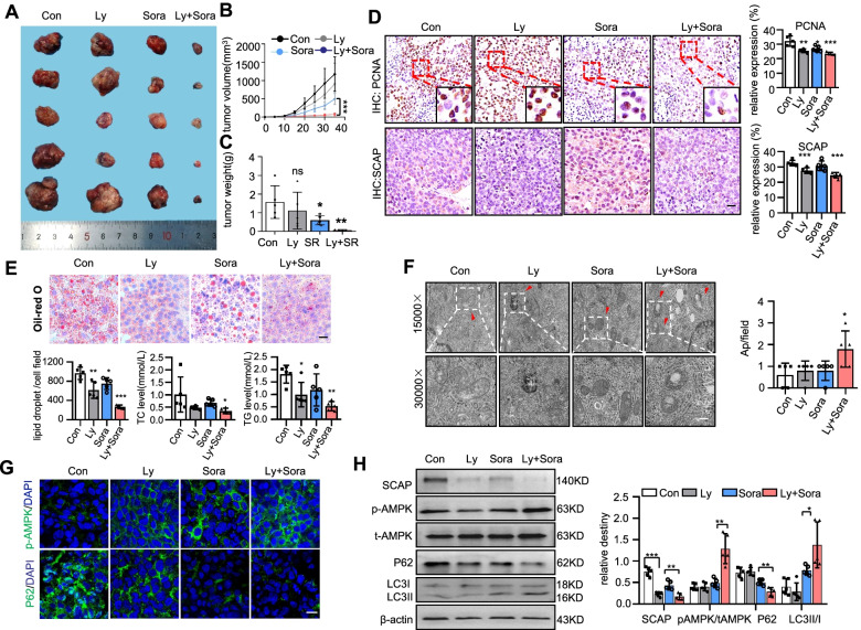
Fig. 6.
SCAP degradation induced by lycorine improves sorafenib resistance in HCC in vivo. A Photographs of subcutaneous tumours after excision (n = 5). B, C Graphs (mean ± SD) showing tumour growth and tumour weight. D PCNA and SCAP staining in tumour tissues (n = 5). E Oil Red O staining in tumour tissues and liver tissues. Bar = 50 μm. The levels of TC and TGs in tumour tissues (n = 5). F Ultrastructural analysis of tumour tissues from each group. The red arrowhead represents autophagic vacuoles (n = 5). Bar = 50 μm. G Representative images of immunofluorescence staining of p-AMPK and P62 proteins in each group (n = 5). Bar = 100 μm. H Immunoblot analysis of SCAP, p-AMPK, t-AMPK, LC3 and P62 protein expression in each group (n = 5). Data are the mean ± SD. *P < 0.05, **P < 0.01, ***P < 0.001. P values were determined by one-way ANOVA in (C), (D), (E) and (F) and repeated-measures ANOVA in (B) and (H)

