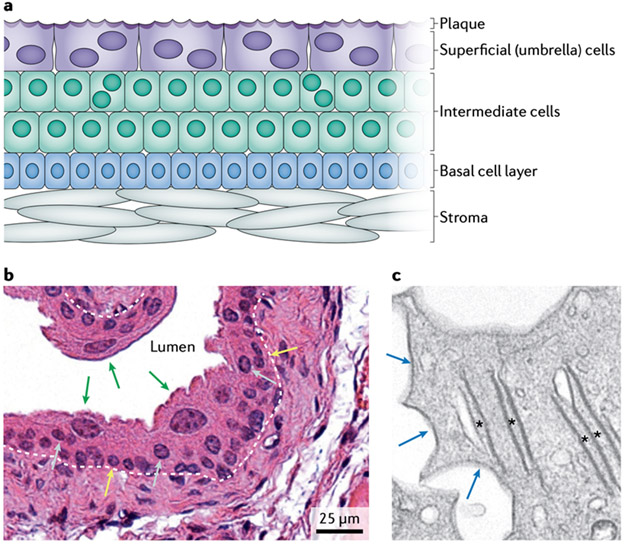
Figure 1. Structural properties of urothelium.
a Urothelium is composed of distinct basal, intermediate and superficial (umbrella) cell populations. Superficial cells secrete an apical membrane plaque composed of uroplakin proteins at the luminal surface. b Haematoxylin and eosin staining demonstrates large, binucleated superficial cells (green arrows), as well as intermediate cells (grey arrows), some of which are binucleated, and basal cells (yellow arrows); ×40 magnification. The urothelial basement membrane is indicated by a white dashed line. c Transmission electron microscopy of superficial cells reveals their unique ultrastructural characteristics, including uroplakin-rich fusiform vesicles (asterisks) and scallop-shaped apical urothelial plaque (blue arrows); ×3,000 magnification.

