Figure 1.

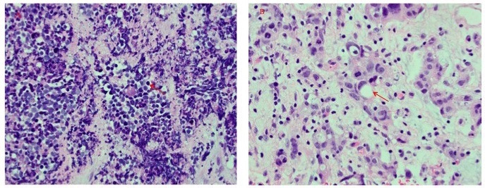
H&E stains of bladder neoplasm (400×). Histologic features of small cell carcinoma (panel (A)) and adenocarcinoma (panel (B)) were both detected. Representative neoplastic cells are labelled (arrows).

H&E stains of bladder neoplasm (400×). Histologic features of small cell carcinoma (panel (A)) and adenocarcinoma (panel (B)) were both detected. Representative neoplastic cells are labelled (arrows).