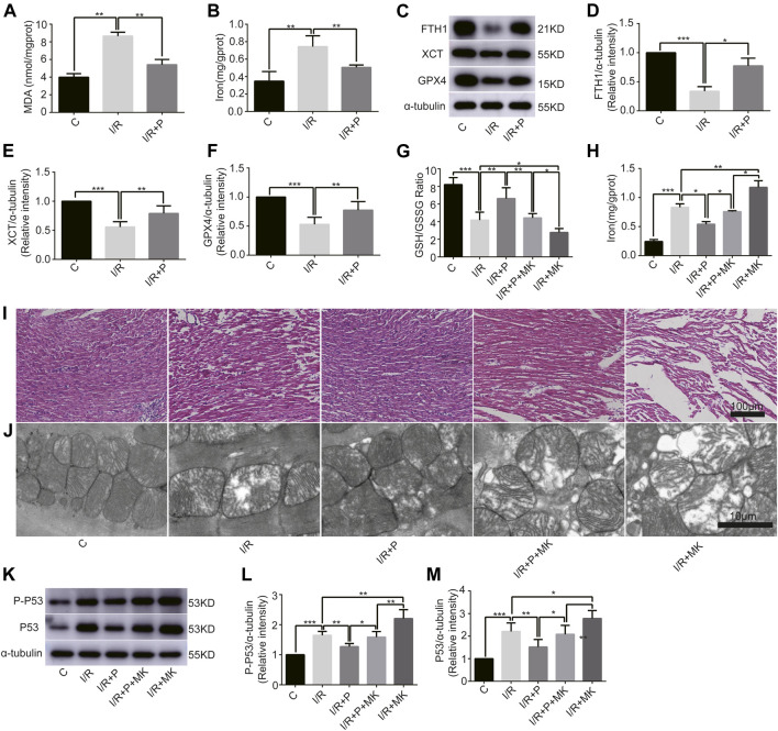
FIGURE 4.
Influence of AKT inhibitor on myocardial ferroptosis with propofol pretreatment. (A–F): Effect of propofol on I/R-induced myocardial MDA, iron, and antioxidant enzymes expressions. (G–H): GSH/GSSG ratio and iron changes in the myocardium. (I) Structural changes in myocardial fibers based on HE staining. Scale bar: 100 μm. (J) Mitochondrial changes under electron microscopy. Scale bar: 10 μm. (K–M): Expression of p53 and p-p53 in myocardium based on the western blot assay. N = 6. Data are expressed as the mean ± SD. Significance was calculated using one-way ANOVA with Tukey’s post hoc test or the t-test. p-values < 0.05 were considered statistically significant. *p < 0.05, **p < 0.01, ***p < 0.001.

