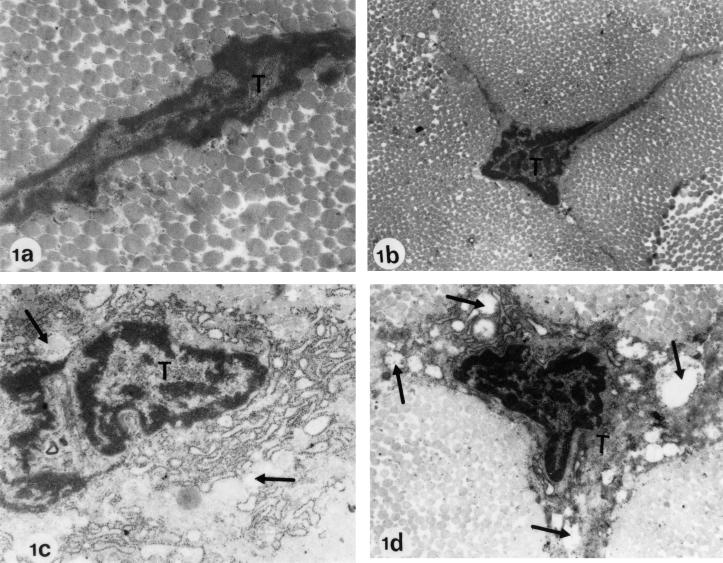
FIG. 1.
Electron microscopy of Achilles tendons from Wistar rats treated at the juvenile stage. (a) Regular diet, no ofloxacin (control; age at investigation, 6 months). (b) Regular diet, no ofloxacin (control; age at investigation, 3 months). Electron micrographs a and b show two typical tenocytes (T) with intact cell organelles. The longitudinal section (a) reveals a regular cell contour. The cross-section (b) shows the typical triangular, irregular shape of tenocytes with wing-like cellular processes. (c) Regular diet plus treatment with one dose of 1,200 mg of ofloxacin/kg (body weight) (age at investigation, day 34). (d) Regular diet plus treatment with one dose of 1,200 mg of ofloxacin/kg (body weight) (age at investigation, 6 months). In panels c and d, pathological alterations after ofloxacin treatment are recognizable. The tenocytes show numerous, partly communicating dilatations and vacuoles in the cytoplasm (arrows). The cell organelles resemble vesicular or piston-like dilatations. Magnification: a, ×34,000; b to d, ×12,750.

