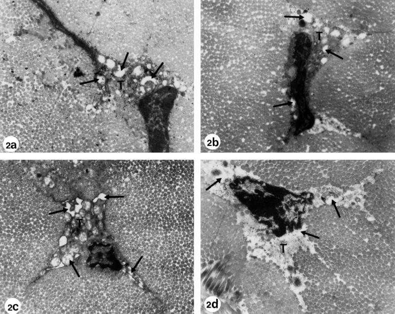
FIG. 2.
Electron microscopy of Achilles tendons from Wistar rats treated at the juvenile stage and fed with a magnesium-deficient diet. (a) Regular diet plus treatment with one dose of 1,200 mg of ofloxacin/kg (body weight) (age at investigation, 3 months). (b) Regular diet plus treatment with one dose of 1,200 mg of ofloxacin/kg (body weight) (age at investigation, 6 months). Electron micrographs a and b show the typical pathological alterations after ofloxacin treatment as described in Fig. 1. The dilatations and vacuoles (arrows) in the cytoplasm of the tenocytes (T) developed due to swellings of the rough endoplasmic reticulum and the mitochondria. Some of the vesicles contain electron-dense material. (c) Magnesium-deficient diet plus treatment with one dose of 1,200 mg of ofloxacin/kg (body weight) (age at investigation, 3 months). (d) Magnesium-deficient diet plus treatment with one dose of 1,200 mg of ofloxacin/kg (body weight) (age at investigation, 6 months). The alterations, such as vacuole formation and dilatation of cell organelles, were more pronounced in rats that had received ofloxacin plus an Mg-deficient diet (panels c and d). The heavily damaged tenocyte in panel d is detached from the surrounding matrix and contains cell debris. Magnification, ×12,750.

