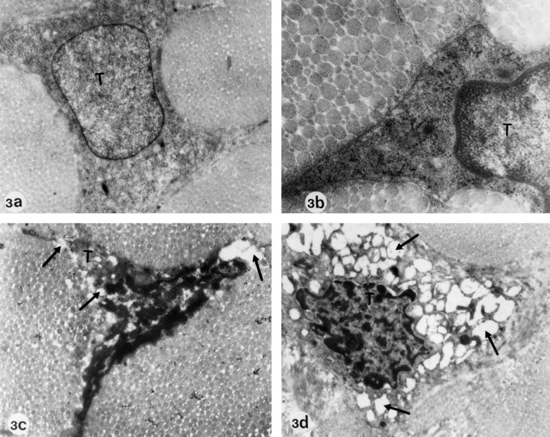
FIG. 3.
Electron microscopy of Achilles tendons from Wistar rats treated at the adult stage. (a and b) Regular diet, no ofloxacin (controls). Magnifications: a, ×26,775; b, ×52,700. Panels a and b show typical tenocytes (T) with a well-demarcated cell surface and regular cell organelles. (c and d) Regular diet plus 10-day treatment with daily doses of 1,200 mg of ofloxacin/kg (body weight). Magnification: ×12,750. Electron micrographs c and d show tenocytes with signs of degeneration similar to those observed in rats that had been treated at the juvenile stage. Vesicle and vacuole formation in the cytoplasm and dilatation and ballooning of cell organelles can be seen (arrows).

