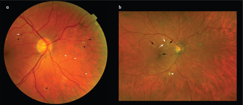
Fig 1.
Colour fundus photography of diabetic retinopathy lesions. a) A photograph of an eye with severe non-proliferative diabetic retinopathy showing dot (white arrowheads) and blot (white arrow) haemorrhages, exudates (black arrowheads) and intraretinal microvascular abnormalities (segments of dilated and tortuous retinal vasculature amid retinal vessels; black arrow). b) A close-up photograph of an eye with diabetic macular oedema showing exudates (black arrows) and microaneurysms (white arrows) at the macula, a cotton wool spot is shown just outside the inferotemporal vascular arcade (white arrowhead).

