Fig 2.

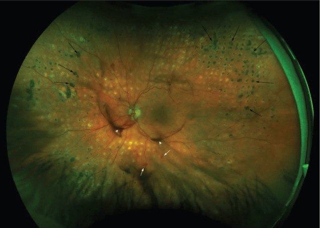
Ultra-widefield fundus photography of an eye with active proliferative diabetic retinopathy showing laser photocoagulation scars (black arrows), new vessels elsewhere (white arrows) and a vitreous haemorrhage (white arrowheads).

Ultra-widefield fundus photography of an eye with active proliferative diabetic retinopathy showing laser photocoagulation scars (black arrows), new vessels elsewhere (white arrows) and a vitreous haemorrhage (white arrowheads).