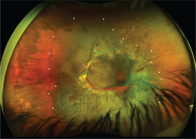
Fig 3.

Ultra-widefield fundus photography of an eye with advanced proliferative diabetic retinopathy showing fibrovascular proliferations (white arrows), new vessels elsewhere (black arrowhead), subhyaloid haemorrhage (black arrow) and tractional retinal detachment (within area of the white arrowheads) involving the macula.
