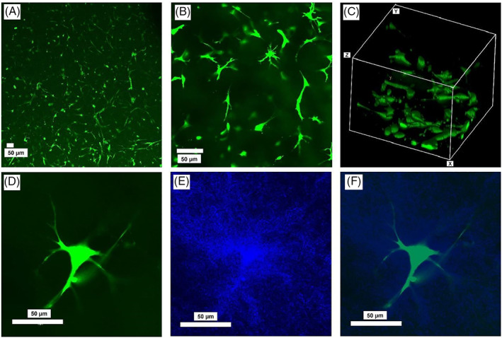
FIGURE 2.

Representative images of AF cells cultured inside type I collagen for 6 days, and then stained with 5‐CFDA‐AM at (A) ×10 and (B) ×20 magnification, and (C) a 3D view, as well as (D) a single AF cell inside collagen, (E) collagen fibers around the cell, and (F) overlayed image of the cell and the collagen fibers (blue). Images were acquired by an inverted confocal laser scanning microscope (Olympus Fluoview 1000; Olympus Life Sciences)
