
FIGURE 9.
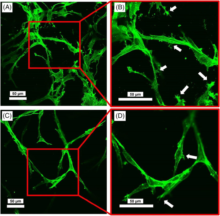
Representative images of cells+collagen constructs 6 days postculture; (A and B) Constructs without LPS treatment at different magnifications (×20 and ×40); (C and D) constructs treated with LPS. Images were acquired by an inverted confocal laser scanning microscope (Olympus Fluoview 1000; Olympus Life Sciences). Arrows indicate the cellular protrusions. Actin filaments formation and organization were not affected; however, cell processes/protrusions appeared to be shorter and less visible for constructs treated with LPS