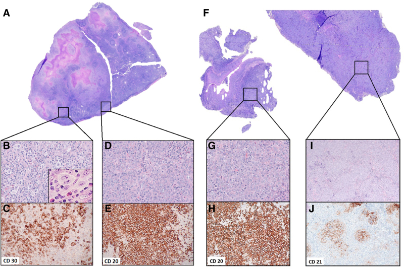
Figure 1.
Morphological features of the composite lymphomas identified in lymph node and tongue biopsies (A) H&E–stained whole slide image of the cervical lymph node biopsy (maximum tissue dimension = 1.8 cm). (B) Lymph node area showing cHL, OM ×40, H&E, inset: high-power view of a representative HRS cell OM ×100 oil immersion. (C) cHL, OM ×20, CD30 highlighting HRS cells. (D) Lymph node area showing DLBCL, OM ×40, H&E. (E) DLBCL, OM ×20, CD20. (F) H&E-stained whole slide image of the tongue mass biopsy (image width = 1.1 cm). (G) Tongue mass area showing DLBCL, OM ×40, H&E. (H) DLBCL, OM ×10, CD20. (I) Tongue mass area showing FOLL3B, OM ×10, H&E. (J) FOLL3B, OM ×10, CD21 highlighting follicular dendritic cell meshwork. cHL = classic Hodgkin lymphoma; DLBCL = diffuse large B-cell lymphomas; FOLL3B = follicular lymphoma grade 3B; H&E = hematoxylin and eosin; HRS = Hodgkin Reed-Sternberg; OM = objective magnification.

