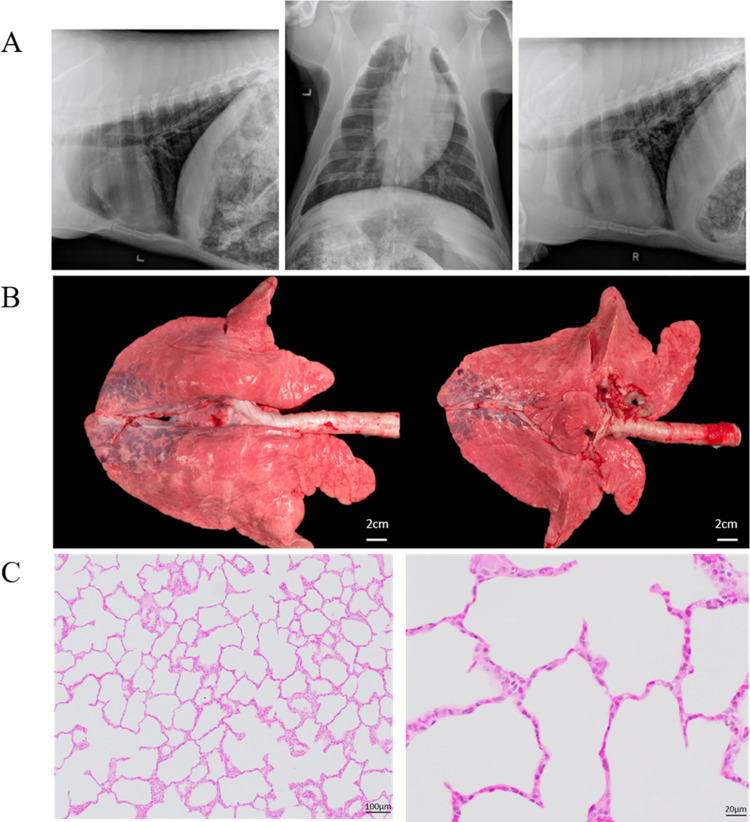
Fig 4. Imaging and histology from the animal experiment.
(A) X-Ray images of the animal’s lungs after the ventilator test showing the (left) left lateral thorax, (middle) VD thorax, and (right) right lateral thorax. (B) Images of the animal’s lungs after the experiment. Note the areas of injured lung in the dependent regions of the lung (C) Histology of the lungs at (left) 10x and (right) 40x magnification. Low-magnification (10x) photomicrograph showing the alveolar duct and the associated alveoli. Note the absence of fluid or proteinaceous debris in the alveoli. High-magnification (40x) photomicrograph of lung after mechanical ventilation demonstrating well preserved alveolar structures. The alveolar septa are very thin and consist of flattened alveolar epithelium (pneumocytes) and delicate capillaries.

