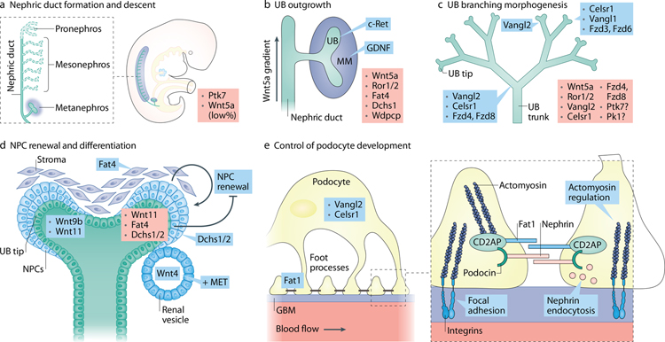
Figure 3: PCP signaling in kidney development.
(A). Nephric/Wolffian duct formation is affected by mutations in certain PCP genes; the known mutated PCP genes are shown in red on the right side of each panel. Expression of PCP genes participating in the discussed process is depicted when known. (B). Outgrowth of ureteric bud from nephric duct is largely controlled by c-Ret (expressed in the ND at the time of UB formation) and its ligand GDNF (expressed in the cells of metanephric mesenchyme. Loss or mutations in several PCP genes lead to abnormal UB outgrowth in both human and mice resulting in renal agenesis or kidney duplication. (C). Ureteric bud branching morphogenesis depends on timely and spatially coordinated changes in cell shape and movements. Mutations in PCP genes affect UB branching, branch shape and branching angles. (D). Nephrogenic progenitor cell renewal and differentiation depend on the crosstalk between stroma and UB tip. Loss of stroma-expressing Fat4 or of NPC-expressing Dchs1/2 leads to NPC expansion. The specific signaling events involving Fat4/Dchs1–2 are unclear. (E). Podocyte foot processes are interdigitated in a precise fashion along the glomerular capillary. Loss of core PCP protein Vangl2 and Celrs1 affects podocyte differentiation, nephrin internalization and glomerular maturation.

