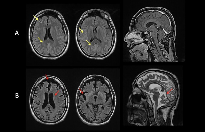
Figure 4.
MRI studies from Patient 1. (A) At age 37 years: T2-FLAIR subcortical hyperintense non-enhancing foci localized to the frontal, temporo-occipital and visual cortices bilaterally with prominent involvement of calcarine cortex (yellow arrows). (B) At age 38 years: severe and diffuse cortical and subcortical atrophy (red arrows).

