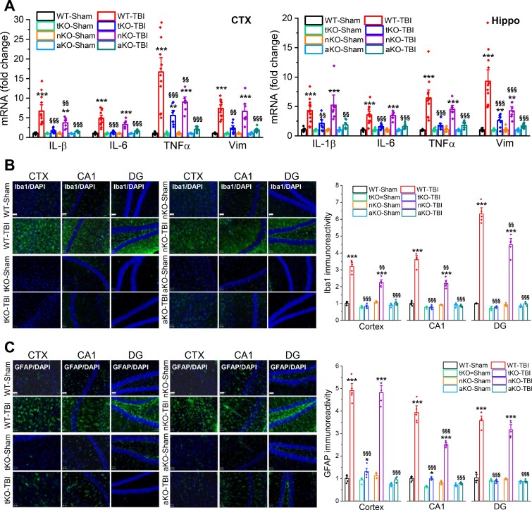
Figure 1.
Inactivation of MAGL in astrocytes suppresses TBI-induced neuroinflammation. (A) TBI-increased expression of proinflammatory factors vimentin and cytokines is alleviated by inactivation of MAGL in astrocytes. The experimental protocols for TBI induction and assessments are provided in Supplementary Fig. 2B. Expression of proinflammatory vimentin (Vim) and cytokines Il1b, Il6 and Tnfa (IL-1β, IL-6, TNFα) in the ipsilateral cortex (CTX) and hippocampus (Hippo) was analysed in wild-type (WT), tKO, nKO and aKO mice 24 h after three impacts. Data are means ± SEM. **P < 0.01, ***P < 0.001 compared with WT-sham, §§P < 0.01, §§§P < 0.001 compared with WT-TBI (ANOVA with Fisher's PLSD post hoc test, n = 7–12 animals/group). (B) Immunoreactivity of Iba1 (microglial marker) in the ipsilateral cortex, hippocampal CA1 and dentate gyrus (DG) was imaged in wild-type, tKO, nKO and aKO mice 30 days after the first TBI. Scale bars = 40 μm. Data are means ± SEM. ***P < 0.001 compared with WT-sham; §§P < 0.01, §§§P < 0.001 compared with WT-TBI (ANOVA with Bonferroni post hoc test, n = 5 animals/group). (C) Immunostaining analysis of reactivity of GFAP (astrocytic marker) in the ipsilateral cortex, CA1 and dentate gyrus of wild-type, tKO, nKO and aKO mice. Scale bars = 40 μm. Data are means ± SEM. **P < 0.01, ***P < 0.001 compared with WT-sham; §§§P < 0.01 compared with WT-TBI (ANOVA with Bonferroni post hoc test, n = 5 animals/group).

