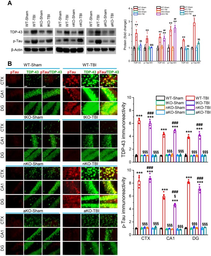
Figure 3.
TBI-induced TDP-43 overproduction and tau phosphorylation are mitigated by inactivation of MAGL in astrocytes, but not in neurons. (A) Western blot analysis of TDP-43, and p-tau-T181 (p-tau) in the hippocampus of wild-type (WT), tKO, nKO and aKO mice 30 days after the first impact. Data are means ± SEM. **P < 0.01 compared with WT-sham; §§P < 0.01, §§§P < 0.001 compared with WT-TBI; ##P < 0.01 compared with nKO-sham (ANOVA with Fisher's PLSD post hoc test, n = 5–7 animals/group). (B) Immunostaining analysis of TDP-43 and p-tau in the brain [cortex (CTX), CA1 and dentate gyrus (DG)] of wild-type, tKO, nKO and aKO mice 30 days after the first injury. Immunoreactivity signals of TDP-43 or p-tau were normalized to WT-sham. Scale bars = 40 μm. ***P < 0.001, compared with WT-sham; §P < 0.05, §§§P < 0.001 compared with WT-TBI; ###P < 0.001 compared with nKO-sham (ANOVA with Bonferroni post hoc test, n = 5 animals/group).

