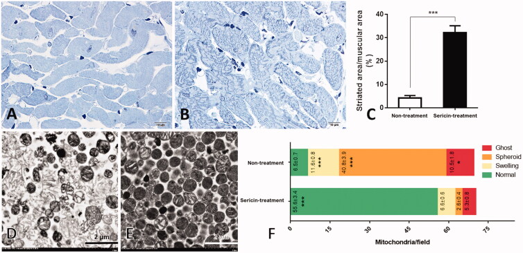
Figure 1.
Histopathology of cardiac muscle and ultrastructure of heart mitochondria: Histopathology of cardiac muscle from hypercholesterolemic rat model was observed through heart non-treated (A) and sericin-treated (B) rats. Percent of striated area per muscular area was demonstrated in the bar graph (C). Electron microscopy was illustrated mitochondrial morphology from heart non-treated (D) and sericin-treated (E) rats. Bar graph (F) represented gold labelling by mean ± SEM comparing each mitochondrial stage; normal (green), swelling (yellow), spheroid (orange), and ghost (red), between non-treated and sericin-treated rats.

