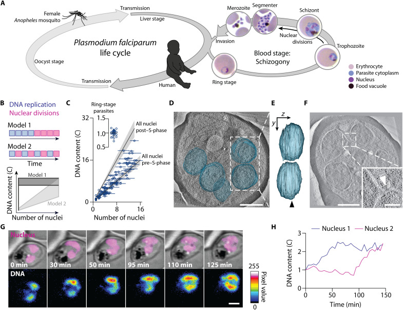
Fig. 1. P. falciparum proliferates through consecutive rounds of asynchronous DNA replications and nuclear divisions.
(A) Scheme of the P. falciparum life cycle, alternating between female Anopheles mosquitoes and humans. Multinucleation occurs in the oocyst, liver, and blood stage. Blood-stage development starts with the ring stage, followed by the trophozoite. Asynchronous nuclear divisions occur in the subsequent schizont stage. At the end of this stage, cellularization occurs and the segmenter is formed, containing nascent daughter cells called merozoites. Egress releases the merozoites that can then infect another erythrocyte. (B) Schematic and predictions of two models proposing the mode of P. falciparum proliferation in the blood stage of infection. (C) Gradual increase of the total DNA content and the number of nuclei of P. falciparum supports model 2. The DNA content was normalized to haploid ring-stage parasites (insert), defined as 1C. Horizontal bars, SD; gray lines, expected DNA contents of parasites with all nuclei before or after S-phase; gray bands, propagated error (SD) of ring-stage measurements. (D) Electron tomogram, overlayed with 3D-segmented inner nuclear membranes (blue); scale bar, 1 μm; see movie S1. (E) Side view of nuclear volumes showed no connection (90° rotation around the y axis); arrowhead, tomogram plane shown in (D). (F) Electron tomogram of connected nuclei; scale bar, 1 μm; inset highlights the connection (arrowhead); scale bar, 250 nm. (G) Time-lapse microscopy of a reporter parasite stained with the DNA dye 5-SiR-Hoechst showed asynchronous DNA replication in sister nuclei; scale bar, 2 μm; see movie S3. (H) Quantification of the DNA content of the nuclei shown in (G).

