
FIGURE 4.
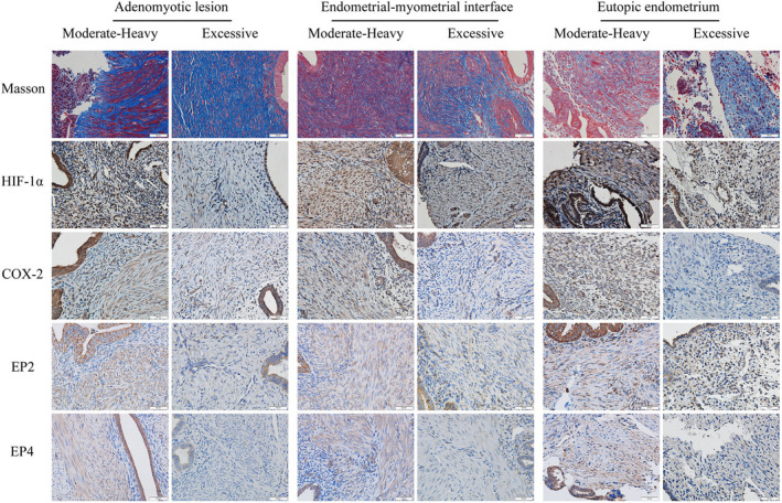
Representative photomicrographs of immunohistochemistry and histochemistry (Masson trichrome) analyses of HIF‐1α, COX‐2, EP2, and EP4, along with the extent of fibrosis in adenomyotic lesions (left panel), their neighboring endometrial–myometrial interface or EMI (middle panel) and eutopic endometrium (right panel) in patients with adenomyosis complaining of moderate–heavy MBL (MHB) and excessive MBL (EXB). The definitions for MHB and EXB are given in the text and Figure 1. Collagen fibers were stained blue, and muscle fibers were red with Masson trichrome staining. Immunoreactivity of HIF‐1α, COX‐2, EP2, and EP4 was observed in both epithelial cells and stromal cells, and HIF‐1α localized both in the cytoplasm and nucleus, while COX‐2 localized in the cell cytoplasm, and EP2 and EP4 in the cell membrane. Magnification: ×400. Scale bar = 50 μm. HIF‐1α indicates hypoxia inducing factor 1α; COX‐2, cyclooxygenase‐2; EP2 and EP4, E‐series of prostaglandin receptors Type 2 and Type 4